Our Community
Sadie’s Sepsis Story

Sadie was very poorly when she was referred to our Emergency and Critical Care (ECC) Service: she had an infection in her womb (known as a pyometra) caused by E. coli bacteria. Pyometra is most common in dogs who have not been spayed, and occurs when bacteria enter the womb during the days of the reproductive cycle when the cervix is open. If the cervix closes after the bacteria have entered, the pus will accumulate inside the womb.
Sadie had been spayed before we met her, however, unbeknownst to everyone, the surgery had not removed the entire womb and a small remnant remained. In Sadie’s case the accumulation of pus had caused her womb to rupture, allowing the E. coli infection to leak into her abdomen, causing sepsis.
Sepsis – also known as blood poisoning – is a life-threatening response to infection due to the body’s immune system overreacting, which causes self-injury and organ dysfunction.
Once Sadie was admitted to our Intensive Care Unit (ICU), the team carried out some point-of-care tests. Point-of-care tests are run in situ, rather than needing to transport the patient to another area of the hospital. They provide fast results, allow the patient to be continually monitored in ICU whilst investigations are performed and reduce patient discomfort and distress.
The results of Sadie’s blood and biochemistry tests were normal. A point-of-care ultrasound scan was also performed, revealing fluid within Sadie’s abdominal cavity surrounding her internal organs. In healthy dogs there should not be any (or only the tiniest amount of) fluid in the abdominal cavity. This fluid was sampled using a small needle and assessed under the microscope in our laboratory.
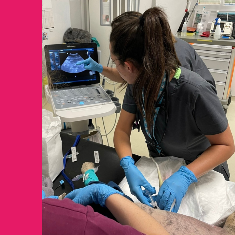
Lorena (ECC Clinician) using point-of-care ultrasound to sample free fluid from the abdomen
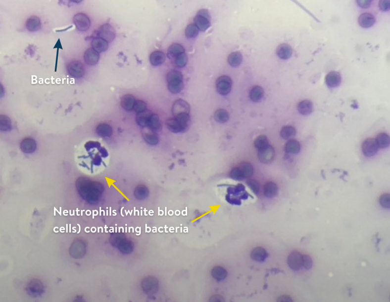
Microscope image showing intracellular bacteria in the fluid within Sadie’s abdominal cavity
The fluid contained a very high number of white blood cells and many of these contained bacteria, a sign of active infection. Sadie was therefore immediately started on intravenous antibiotics.
The point of care ultrasound scan raised concerns that other organs were affected; the urinary bladder, right kidney and ureter appeared abnormal. The team needed more information, and so they performed a CT scan under general anaesthesia. The CT scan revealed a ‘stump’ pyometra (infection of a remnant of the womb) and confirmed the presence of abdominal free fluid as well as severe inflammation of the abdominal cavity (peritonitis).
Sadie was urgently transferred to theatre and underwent an exploratory laparotomy (surgical opening of the abdomen). Our surgical team found that Sadie had severe peritonitis as well as severe adhesions between her bladder, ureters and colon.
In healthy dogs, the abdominal organs glide against each other, but in patients with peritonitis, strong fibrous adhesions develop which causes the organs to stick together. These adhesions are not only painful, but they also interfere with normal organ function and health.
It took a significant amount of time for the surgical team to gently break down the adhesions and separate Sadie’s organs. A small section of Sadie’s bladder had to be completely removed (partial cystectomy). The infected remnant of her womb was also removed, and warm sterile saline was used to flush and clean the abdominal cavity before closing.
Sadie was taken back to our ICU to recover from surgery and general anaesthesia. She was carefully monitored for any complications resulting from such a significant surgery. Over the days that followed, she improved little by little, becoming brighter with an increasingly healthy appetite. When she was well enough, Sadie was discharged to continue her recovery at home with her family.
Thank you for reading Sadie’s story. For regular news and insights from The Ralph team, as well as stories about our lovely patients, be sure to check out our Instagram, Facebook and LinkedIn.
Take care,
Team Ralph 🐾