Our Community
The Ralpher – December 2019 Edition
This newsletter is intended for a veterinary audience and contains clinical photographs
Hello!
Welcome to our last edition of 2019! As we strive to deliver a quality service to our vet community, we have welcomed a number of new faces to our team of specialists (read all about it below). Our Oncology Service launched in October with Stefano Zago, and our new Cardiology Diplomate, Luca Ferasin, shares more about his love of cardiology. This month’s featured patient stories include Frankie’s rare cause of urinary incontinence, and the use of medical intervention for Baylie’s seizures. Our monthly free CPD events will continue in 2020, with video and audio recordings from this year’s events available on demand via our website – check out our Library + Learning page. Wishing you a cosy winter season and a warm welcome to 2020!
Take care,
Team Ralph
In this month’s edition…
Spotlight on…
Meet our new Oncologist Stefano and learn about our new Oncology Service
Cardiology in focus…
We get to know our newest Ralpher and Cardiology Diplomate Luca
Tales from the clinical floor:
This month we hear Baylie and Frankie’s stories
Team announcements – A trio of Diplomates!
We are delighted to welcome Luca Ferasin, Stefanie Mitze and Veerle Volckaert to Team Ralph.
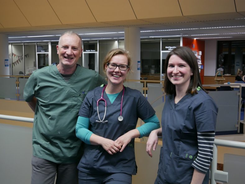
From left to right: Luca, Stefanie, Veerle
Luca Ferasin DVM PhD CertVC PGCert(HE) DipECVIM-CA (Cardiology) GPCert(B&PS) FRCVS European & RCVS Specialist in Veterinary Cardiology brings a wealth of experience to the team. Luca has been a specialist for over 15 years and was recently awarded a Fellowship of the Royal College of Veterinary Surgeons for his Meritorious Contributions to Clinical Practice. Luca is an active researcher and has also vastly contributed to the veterinary literature with articles, abstracts, and book chapters, including the chapter on coughing in the latest edition of Ettinger’s Textbook of Veterinary Internal Medicine. Luca joins fellow Cardiologist Heidi Ferasin to deliver a specialist-led Cardiology Service.
Stefanie Mitze DVM DipECVIM-CA (Internal Medicine) MRCVS joins our Internal Medicine Service. Stefanie graduated in 2010 from the Szent István University in Budapest, Hungary. Since then she has worked in several multidisciplinary referral centres in Germany, including the University of Munich. Stefanie completed her residency in a private clinic in Munich (Tierklinik Haar) from 2014-2017. She has undertaken an externship at the University of Bern in Switzerland and the University of Utrecht in the Netherlands, as well as at the Royal Veterinary College.
UPDATE November 2020: Stef has left Team Ralph to pursue other veterinary projects. We wish her all the very best!
Veerle Volckaert DVM MRCVS PhD DipECVDI joins our Diagnostic Imaging Service, an integral part of the hospital. Veerle graduated from Ghent University as a Master in Veterinary Medicine in 2010. She continued working at the university doing an internship, followed by a residency under the European College of Veterinary Diagnostic Imaging (ECVDI). Veerle received the diploma of the ECVDI in 2015 and finished her PhD in nuclear medicine at Ghent University in 2016. Veerle joins us having worked in a small animal referral setting for a number of years.
Spotlight on Oncology
Our new Oncology Service launched in October with Oncologist Stefano Zago. Whilst we had always planned to offer an Oncology Service, this launch was a little earlier than we had anticipated. Over the past eight months, this had been one area of veterinary medicine where we have not been able to serve our patients, their carers, or our referral community. This did not sit well with us. Serendipity being what it is, our path crossed with Stefano’s and we were able to extend our services.
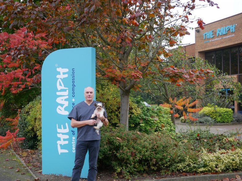
Stefano and Dempsey
Stefano joins us with oodles of experience in caring for patients with cancer, and an interesting history of working with animals across the globe. He was born and raised in Italy and studied in Parma, graduating with Honours. Following graduation, Stefano attended military service with the Presidential Horse Guards as a Veterinary Officer. Following a Master’s degree in Tropical Veterinary Medicine at the University of Edinburgh, Stefano worked with the United Nations in Somalia, Malaysia and Guyana in wildlife management, dealing mainly with rhinos, orangutans and crocodiles. Stefano returned to the UK to care for companion animals and completed a Master’s in Clinical Oncology at the University of Birmingham. He has worked in a number of busy referral practices since 2015, managing and treating small animal oncological cases. Stefano enjoys all aspects of medical oncology and has a specialist interest in lymphoma, haemangiosarcoma and metronomic chemotherapy.
Stefano leads the Oncology Service which provides diagnoses, treatment, and care for dogs and cats with cancer. We provide the following medical therapies:
– Conventional chemotherapy: uses the same drugs as are used in humans with cancer. But we reduce the dosages, simplify the protocols and extend the treatment intervals. This allows our patients to go through treatment usually with little to no side effects.
– Metronomic chemotherapy: the administration of a small, usually daily, dose of drugs to inhibit the growth of the tumour.
– Targeted molecular therapies: tyrosine kinase inhibitors differ from conventional chemotherapy. They work by binding to specific receptors on cancerous cells. This prevents the cancer cells from dividing and growing which slows or stops the growth of the tumour.
– Immunotherapy: uses the body’s immune system to fight the cancer. We offer melanoma and lymphoma vaccines as treatments for these types of cancer.
We understand a diagnosis of cancer can be a very distressing and confusing time for everyone involved. That’s why we feel it is important to support practices and members of the veterinary community to help their clients and their pets.
For any oncology-related queries or advice, please get in touch with Stefano. We are always happy to help. Drop us a line at [email protected] Get in touch!
Cardiology in focus
Luca Ferasin, Cardiology Diplomate and latest Ralpher to join the team, shares his words of wisdom and what keeps inspiring him along his journey to Fellowship and beyond…
What inspired you to become a Cardiology Specialist?
I wanted to be a cardiologist since I was a young child, having been particularly inspired by my uncle who was a very talented and compassionate human cardiologist. At the age of just 15, I was spending several days shadowing a friend who was a cardiology nurse in a human hospital and was completely captivated by this fascinating clinical activity. At the same time, I did not want to neglect my true love and passion for animals, so I ended up becoming a veterinary cardiologist.
What exciting developments are there from the latest in cardiology research?
Cardiology is probably one of the fastest growing disciplines in veterinary medicine and I have been very fortunate to be involved in advanced clinical research over the years. I have been transferring this knowledge to my daily clinical activity to provide the highest standard of care and best quality of life to my patients.
What is the most common cardiology condition you treat, and why is it so common?
Mitral valve disease in dogs is certainly the most common condition that small animal cardiologists see on a daily basis. It is an age-related disease and it is becoming more common due to the extended life expectancy of our pets.
What is your top-tip for primary care clinicians?
I always stress the importance of an accurate physical examination. Indeed, clinical findings, in combination with a thorough evaluation of the pet’s history, can provide 90% of the final diagnosis and prevent the need for expensive diagnostic tests.
What does “Be more Ralph” mean to you?
The Ralph’s core values are the very same values that have driven my long career. The fact that the ethos is truly respected at The Ralph and implemented at every level adds extra value to my job.
Luca and Luna
Luca joins Heidi Ferasin in our specialist-led Cardiology Service providing diagnoses, management and treatment for patients with a range of conditions. For any questions or advice, you can email Heidi and Luca by contacting [email protected]
Tales from the clinical floor
Baylie
A visit to the groomers doesn’t usually involve frothing at the mouth, but that is sadly what happened to Baylie, a 5-month old miniature Poodle-cross, when she had a seizure in the groomer’s bath tub.
Two weeks later another seizure occurred, on dry land this time: Baylie was answering the call of nature when she went rigid, fell over and frothed at the mouth. Just over a week later she had another. Baylie was thoroughly assessed by her local vet, and metabolic causes of seizures were ruled out by comprehensive blood tests.
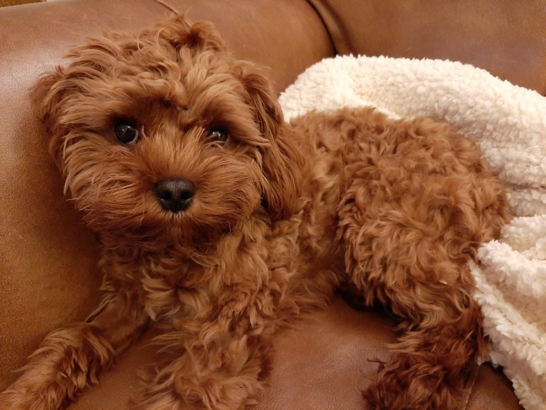
Baylie at home
Baylie was referred to Sue Fitzmaurice, Head of Neurology + Neurosurgery, for further investigation. The only abnormality on the neurological exam was the menace response, which was absent in the right and equivocally present on the left. At 5-months of age, Baylie was old enough to have learnt the menace response, and her alertness suggested that she should have been paying attention. No other deficits were found. The clinical sign of seizures localised the lesion, as neurologists like to say, to the cerebrum. The absence or reduction of a menace response with normal PLR can also suggest a cerebral localisation. An MRI of Baylie’s brain was recommended.
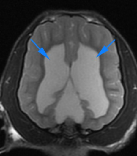
Dorsal T2W image showing the bilateral lateral ventricles dilation
The MRI of Baylie’s brain found enlargement of the lateral and third ventricles and also of the CSF-filled area above the quadrigeminal (tectal) plate of the midbrain. Enlargement of the quadrigeminal cistern has been described in dogs as both a cause of clinical deficits (ataxia) and as a subclinical finding. It appears to be rare: 2.2% of MRI studies found supracollicular fluid accumulations.*
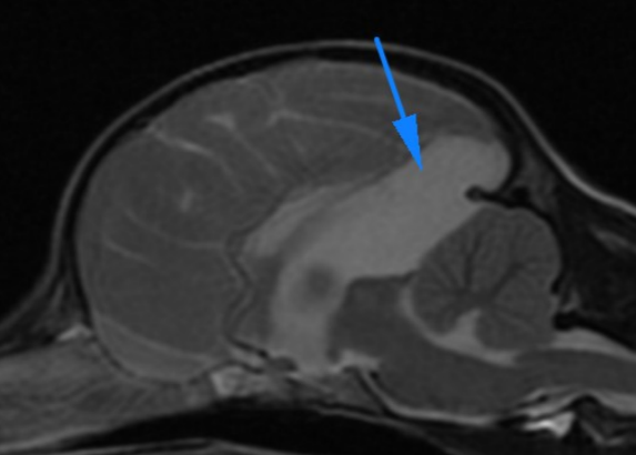
Sagittal image showing the cerebrospinal fluid like accumulation in the supracollicular area
The normal quadrigeminal cistern is separated from the third ventricle by a thin membrane. The radiologist thought that this membrane was missing in Baylie. The fluid accumulation in the quadrigeminal cistern compressed the rostral margin of the cerebellum and the caudal margin of the occipital lobes. Baylie was started on phenobarbitone to control the seizures. Her menace response was normal bilaterally on re-exam, so the variations were thought to be due to her inattention rather than the hydrocephalus. The cause of Baylie’s seizures is either the hydrocephalus, or the compression of her occipital lobes, or, given her breed, idiopathic epilepsy. As she remains normal between her seizures, no surgical intervention is planned. Her behaviour at home will be the best measure of her brain function, and so the 6-monthly check ups at her local vet will be very important over the next few years while we assess the significance of her brain malformations.
Do you have a neurology-related question, or are you looking for some advice? Then drop us a line, we are always happy to help. You can email [email protected] or call 01628 308330
*Reference: Vet Radiol Ultrasound. May 2016;57(3): 259-68.
Frankie
Frankie is a 6-month old Dachshund who recently presented to our Soft Tissue Surgery Service for investigations into her urinary incontinence. The carers reported that she was leaking urine at night as well as dribbling urine during the day.
Haematology and biochemistry were unremarkable. Urinalysis demonstrated a urinary tract infection. Frankie was sedated and an abdominal ultrasound was performed. Ultrasonography demonstrated a large, thin-walled, cystic structure just caudal and to the left of the bladder. This fluid-filled structure resembled an additional bladder. To clarify the abnormal urinary tract further an intravenous urogram was performed with CT. The CT scan demonstrated a dilated left ureter with mild hydronephrosis. In addition, both the fluid-filled structure just caudal to the bladder and the bladder itself filled with contrast to give the appearance of a second bladder. The termination of the left ureter could not be confirmed but did not appear to be entering the bladder.
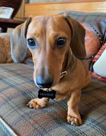
Frankie at home
The “double bladder” sign on imaging is consistent with a ureterocoele. A ureterocoele is a rare congenital defect that results in the dilation of the distal ureter. This dilation can occur within the bladder lumen or as the ureter enters the bladder neck, and can lead to recurrent urinary tract infections, urinary incontinence as well as ureteral obstruction. Ureterocoeles commonly occur in conjunction with ectopic ureters and congenital urethral sphincter muscle incompetence, and therefore management of these complex cases can be challenging.
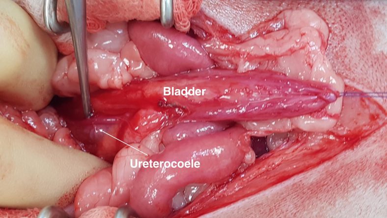
Ureterocoele visible within the wall of the bladder neck and urethra
A ventral celiotomy was performed to evaluate the urinary tract further. The left ureter was clearly dilated (approximately 3x the size of a normal ureter). A ventral cystotomy was performed. The ureterocoele was visible within the wall of the bladder neck and urethra. There was no evidence of the left ureter opening into the bladder, and when the ureterocoele was incised and a urinary catheter passed retrograde, it was apparent there was a concurrent left intramural ectopic ureter opening into the vagina. The distal left ureter was ligated and transected to allow reimplantation of the distal ureter into the apex of the bladder, a procedure called a neoureterocystotomy.
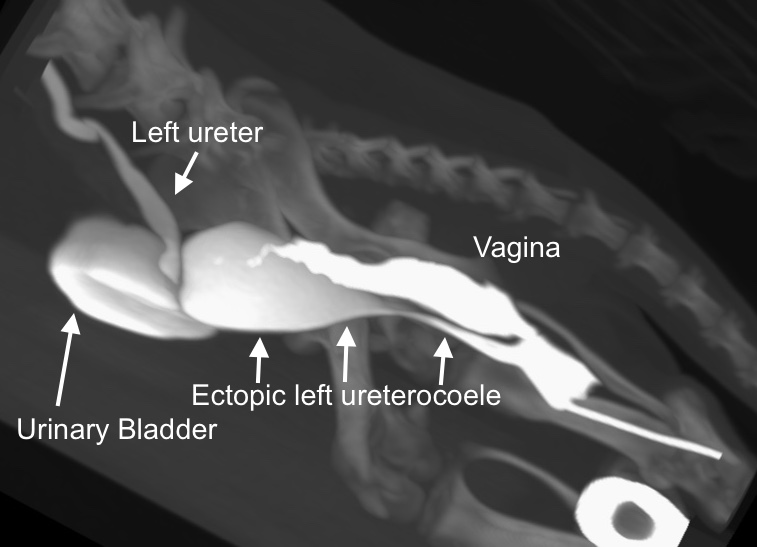
3D image of the lower urinary tract after contrast administration (CT intravenous urogram and retrograde vagino-urethro-cystogram). Contrast is visible in the dilated left ectopic ureter, in the urinary bladder, and in the ectopic ureterocoele. There is also contrast in the vagina and a small amount in the uterus
Frankie recovered really well from the surgery and after a short hospitalisation stay, she continued her recovery under the watchful eyes of her carers. Urine microbiology demonstrated a concurrent urinary tract infection, so a short course of antibiotics was prescribed. The carer reports resolution of the urinary incontinence during the day, however some leaking of urine still occurs at night. The success for resolution of urinary incontinence following surgery for ectopic ureters and ureterocoeles is 50-60%. The most likely reason for this low success rate is concurrent congenital urethral sphincter muscle incompetence, as suspected in Frankie’s case. In cases of congenital urethral sphincter muscle incompetence, it is advisable to not neuter bitches until after their first season as the signs can resolve/improve following oestrus. However if the urinary incontinence does not resolve/improve then medical management with phenylpropanolamine/ oestrogens, or surgery with a urethropexy, cystopexy, colposuspension or artificial urethral sphincter should be considered.
For any surgery-related queries or advice, please get in touch. You can email the team on [email protected] or call 01628 308330.
Thank you for taking the time to read our news and stories from inside the hospital. Stay tuned for news on our upcoming events plus more stories from inside the hospital.
Take care,
Team Ralph