Our Community
Ghost’s story
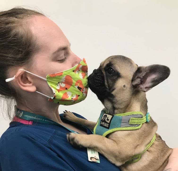
Ghost with our Cardiology Nurse, Amy
Following detection of a heart murmur at her primary care vet and a visit to our Puppy/Kitten Heart Murmur Clinic to investigate this, Ghost – who is under the care of Dogs Trust – was diagnosed with pulmonic stenosis.
The pulmonic valve sits between the right ventricle of the heart and the pulmonary artery, which is the major blood vessel that carries blood from the heart to the lungs. Pulmonic stenosis is where the blood flow between the heart and lungs is obstructed by a malformed pulmonic valve.
This is one of the most common congenital defects in dogs but is particularly prevalent in some brachycephalic (short-nosed), spaniel and terrier breeds. In young dogs, the only sign of this condition may be the presence of a heart murmur. However, some dogs may show signs of exercise intolerance, breathing difficulties and syncope (fainting). If a heart murmur is detected in a puppy or symptomatic patient, referral for assessment by a Cardiologist may be recommended.
Our Cardiologists used an echocardiogram (ultrasound scan of the heart) to diagnose Ghost’s cardiac disorder and to assess the amount of pressure the condition was putting on her heart. This is measured in mmHg (units of pressure) and is calculated by measuring the speed of blood flowing across the pulmonic valve from the right ventricle into the pulmonary artery. Normal pressure across the pulmonic valve is usually between 15-25 mmHg and Ghost’s was measured at approximately 70 mmHg with severe thickening of the right ventricular wall due to the high pressure!
Balloon valvuloplasty (BVP) is currently the best available treatment for Ghost’s condition. This minimally-invasive procedure involves the insertion of a catheter through the jugular vein in the neck which, under guidance with fluoroscopy (real-time X-rays), is passed inside the heart and into the pulmonary artery. The catheter has a sausage-shaped balloon on the end, which is placed across the narrowed pulmonic valve and, when inflated, widens the opening of the valve, therefore reducing pressure and increasing blood flow between the heart and lungs.
Ghost’s surgery was a success and she made a rapid recovery from anaesthesia. After close overnight monitoring by our wonderful team of nurses, Ghost was discharged the following day.
During her initial recovery period, it was important for Ghost to only have gentle lead walks. At her most recent check up, an echocardiogram showed that the pressure across Ghost’s pulmonic valve had reduced from 70 mmHg to 30 mmHg. This means she was gradually able to increase to her normal level of activity – go Ghost go!
Thank you for reading Ghost’s story. Check out Our Community page for more patient stories, and our Cardiology page if you’d like to know more about the service and our team.
Take care,
Team Ralph.