Our Community
Advanced spinal surgery in 2.4kg Dotty
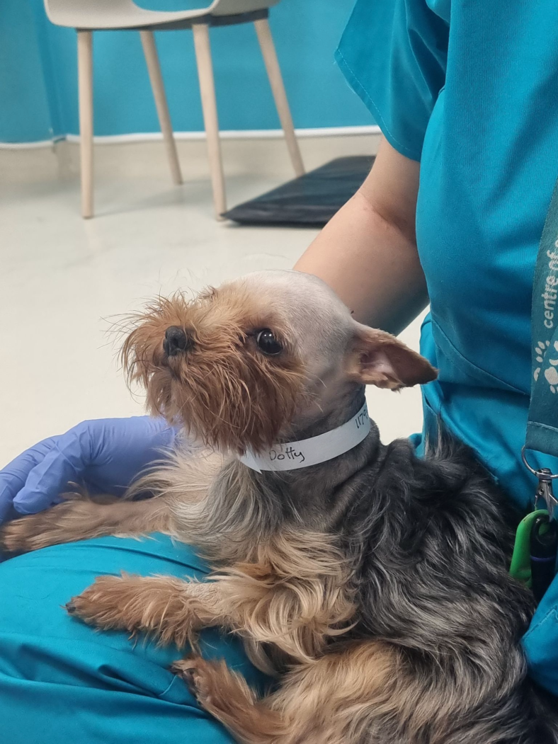
2.4kg Yorkshire Terrier, Dotty, was referred to The Ralph’s Neurology and Neurosurgery team following sudden paralysis in all four of her legs. A full neurological examination, including an MRI and CT scan, confirmed the suspected diagnosis of Atlanto-Axial subluxation.
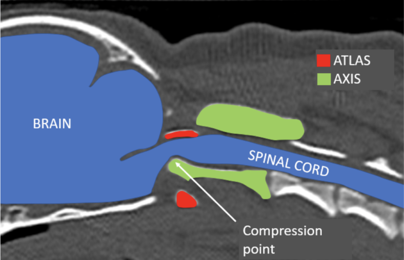
Atlanto-Axial (AA) subluxation is a condition most common in “toy breeds” where the first two vertebrae of the neck (the atlas and the axis) subluxate (misalign), which traumatises and compresses the spinal cord. This condition can be congenital (present since birth) or traumatic (caused by an external source). AA subluxation can present in many ways, from just episodic pain to sudden paralysis, respiratory difficulties and even sudden death.
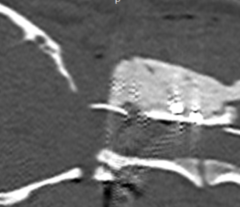
Below you can see an annotated CT scan of Dotty’s spine before surgery:
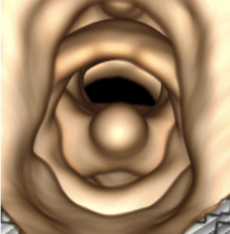
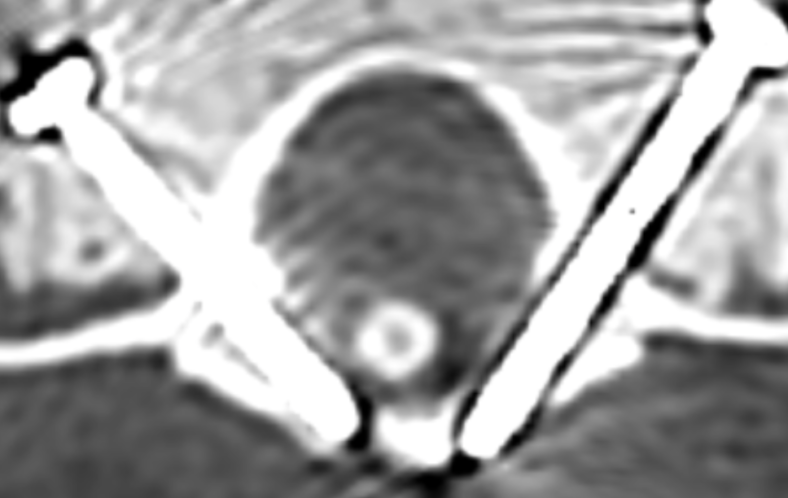
And a 3D reconstruction of Dotty’s spinal canal. See the round tip of the axis in the centre of the vertebral canal, leaving very little space for the spinal cord to run:
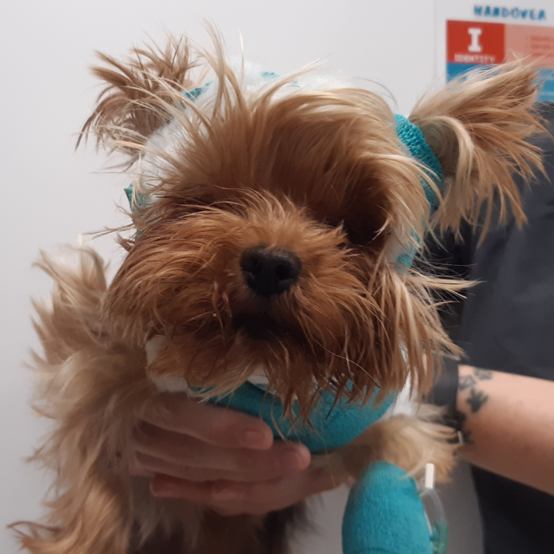
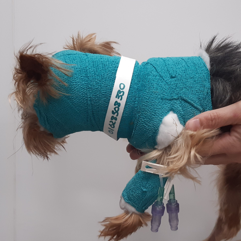
Dotty had to wear a protective bandage to limit her neck movements while waiting for her surgery (see below). Some cases can be managed conservatively (i.e. without surgery); however, this means that the dog has to wear this protective bandage for several weeks, with regular bandage changes to prevent skin lesions. The time needed to recover can be longer than with surgery and the risks of further subluxation will still exist in the future, which means physical activity must be limited in the long run. That’s why our Neurology team recommended surgery as the best treatment option for Dotty’s quality of life both in the short term and long term.
Our Head of Neurology and Neurosurgery, Lorenzo, operated on Dotty with a “novel dorsal AA stabilisation technique“. The technique was designed and published last year by Dr Leblond and his colleagues, but modified by our team in collaboration with Vet 3D by combining it with the use of 3D printed guides which maximise the precision of screw placement. The guides are applied on the dog’s vertebrae during surgery and will guide the drill to the safest and most effective part of the bone to place a screw.
Check out the process in images: 3D printed models, an insight into the surgery and post-operative CT scans showing normal alignment of the atlas and axis, with the screws positioned perfectly and the spinal cord decompressed…
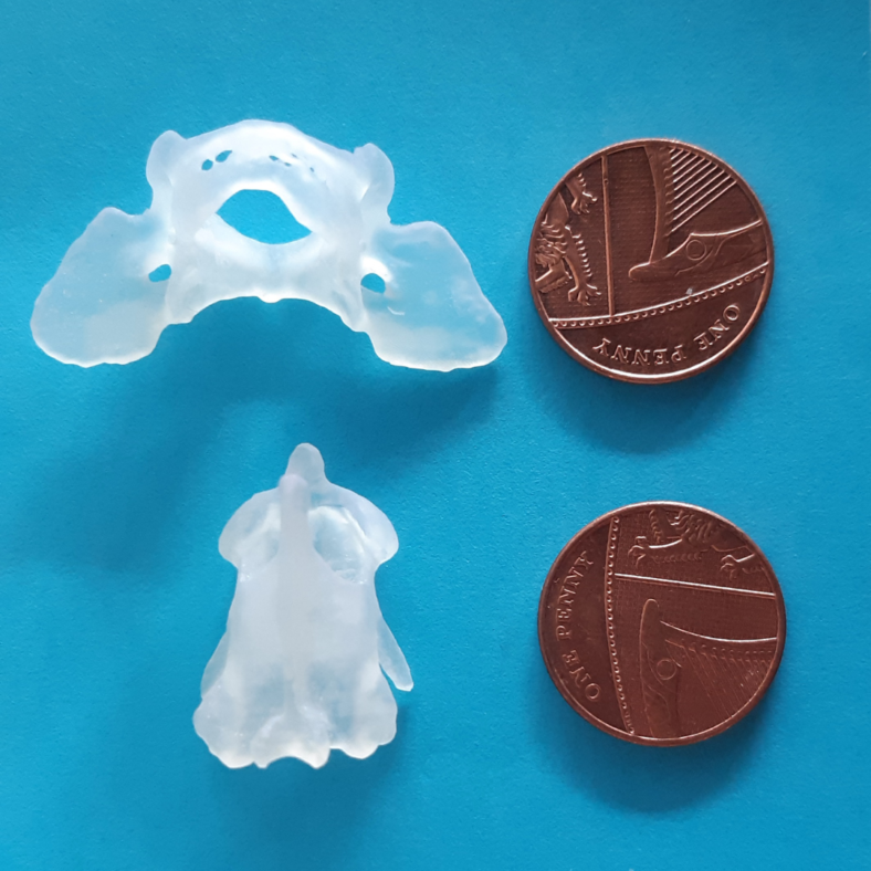
3D printed to-scale models of Dotty’s atlas and axis, with pennies for size-comparison
Watch Lorenzo realigning the axis to its correct position during a dorsal Atlanto-Axial stabilisation surgery:
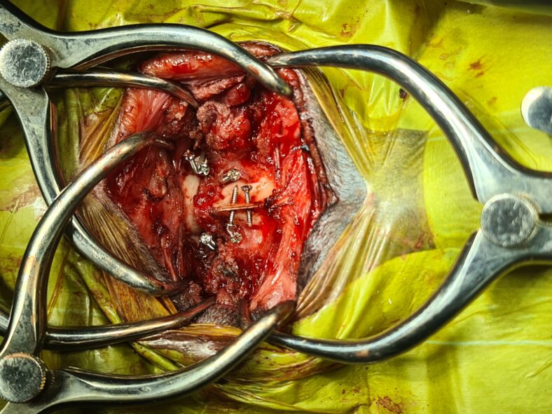
Intraoperative picture with all screws placed, ready to place the surgical cement
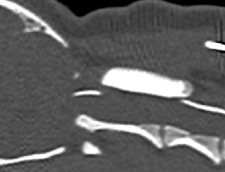
Comparison of the pre-op and post op CT, showing the correct alignment of the atlas and axis after surgery:
Pre-op CT scan
Post-op CT scan showing correct alignment of the atlas and axis
Detail of the precise placement of the screws in the atlas with the use of 3D printed guides
The result was beyond belief! Dotty started walking again just two days after surgery, and even tried to run (although it was too soon to allow her to!)
Having recovered well from her general anaesthetic and surgery, Dotty was discharged and able to continue the rest of her recovery journey at home with her family. Dotty’s family needed to rest her in a crate, and avoid Dotty getting hyperactive, with very short incremental walks on a lead and harness.
Lorenzo said, “Dorsal stabilisation techniques (i.e. from the top of the neck) present very many advantages compared to the most classically used ventral techniques (i.e. from underneath the neck). Not only do they better respect the biomechanical principles of fracture/luxation stabilisation, but also, and most importantly, they avoid dissections close to extremely important and delicate organs such as the larynx, the pharynx, the oesophagus, and very important nerves and vessels, minimising the risk of serious complications related to post-operative dysfunction of these structures.
Dorsal to the spine indeed there are only muscles, providing much more space to place the surgical cement that joins the screws together, which can also cause mechanical compression or irritation to the above listed structures when placed ventrally. The problem of dorsal techniques has always been that the constructs previously described appear to be pretty weak compared to the ventral techniques, and this is where Dr Leblond technique changed the game, having the merit to identify safe and pretty generous bone corridors to introduce the screws. I was lucky enough to work with Dr Leblond last year and learn this technique directly from him.

The unique modification that we made here at The Ralph, in collaboration with Vet 3D, was to combine this technique with the use of 3D printed customised guides that can maximise the precision in placing screws in each single case. We must not forget that most of these dogs weigh around 2kg and we are talking about inserting 1.5mm screws in bones that are 2.3-2.5 mm in thickness, where the margin for error is close to zero.”
Here is a short video of Dotty at her most recent check-up – one month after her surgery!
We are so pleased for Dotty and her family, and proud of all involved in helping Dotty get back on her tiny feet again.
Thank you for reading Dotty’s story. For more patient stories, check out our Instagram, Facebook and LinkedIn!
Take care,
Team Ralph 🐾
P.S. Hey VETS – did you know we’re holding a four-part Neurology CPD series? Check out our events page for all you need to know!